Check out our interactive online atlas!
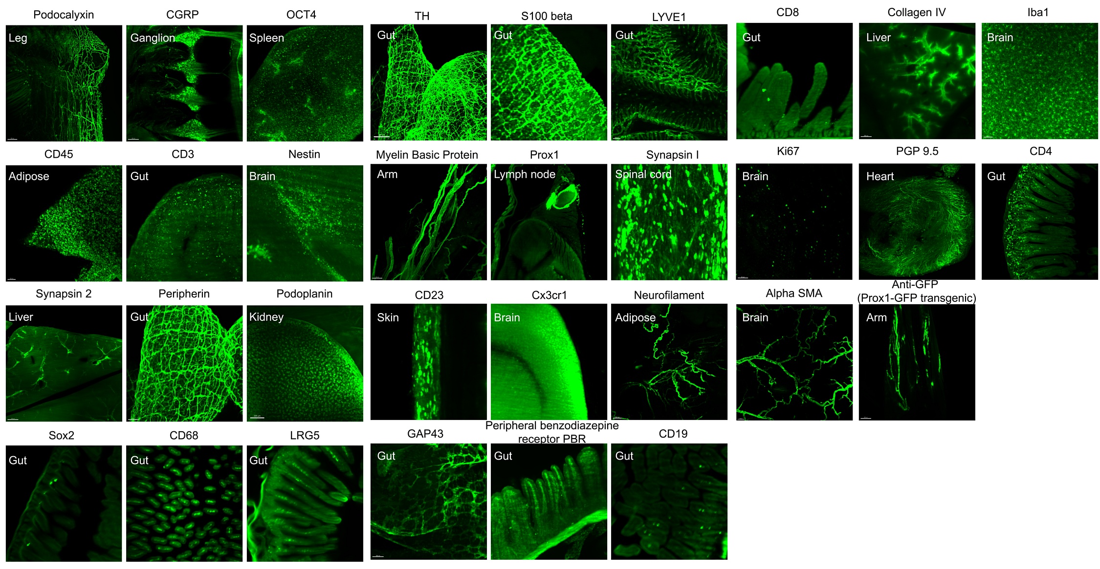
Antibody validation, click on image for full resolution (24MB) or download the list in PDF

Supplementary content
Movie S1
3D reconstruction of a mouse labeled with PGP 9.5 and imaged with light-sheet microscopy. Different PGP 9.5 innervation regions (green) are visualized with high contrast over background (gray).
Movie S2
3D view of PGP 9.5 positive peripheral nerve in mouse heart showing in magenta.
Movie S3
3D visualization of sympathetic nerve in mouse spleen labeled with tyrosine hydroxylase (TH) in magenta.
Movie S4
3D visualization of mouse liver and gallbladder innervated with PGP 9.5 positive peripheral nerves in magenta.
Movie S5
3D annotation of mouse intestinal innervation with TH positive nerve in magenta. Grid-like lattice structures can be seen in the intestinal wall.
Movie S6
3D annotation of neuronal connections in multiple organs (kidney, spleen, liver, and intestine) labeled with PGP 9.5 in green.
Movie S7
3D visualization of the entire lymphatic vessels in the whole mouse. The lymphatic vessels of a 4-weekold
mouse were labeled with LYVE1 in yellow.
Movie S8
3D visualization of the hind limb of a mouse with LYVE1 lymphatic vessel labeling in yellow. Fine
details of the lymphatic vessels and lymph nodes are shown.
Movie S9
3D visualization of mouse kidney with LYVE1 lymphatic vessels labeled in yellow.
Movie S10
3D annotation of mouse stomach with LYVE1 lymphatic vessels highlighted in yellow. The fine details of the lymphatic vessels are clearly visible throughout the scan.
Movie S11
3D annotation of mouse intestine lymphatic vessels with yellow color labeled by LYVE1 and scanned with light-sheet microscopy. Fine details of the lymphatic vessels can be seen in the intestine and a lymph node is located adjacent to the intestine.
Movie S12
3D view of the intestinal wall of a mouse with LYVE1 lymphatic vessels labeled in yellow.
Movie S13
wildDISCO staining of PROX1 lymphatic vessels in green and arterial staining of alpha-SMA in red, revealing lymphatic vessels penetrated in mouse cerebral cortex.
Movie S14
3D illustration showing wildDISCO staining of LYVE1 in yellow and podoplanin in magenta to visualize lymphatic capillaries covered on the surface of the mouse brain, and also entering the brain parenchyma around thalamus.
Movie S15
3D illustration showing innervation of intestinal lymph nodes by sympathetic neurons using TH staining to label sympathetic nerves in magenta and CD45 staining to label immune cells in green.
Movie S16
Representative 3D reconstructions of immune cells on the intestine neurons. CD45 is stained in green for immune cell distribution and TH stained in magenta for sympathetic nerves.
Movie S17
3D reconstruction of a TH and LYVE1 labeled mouse by light sheet microscopy. Different regions of sympathetic nerve innervation in green and lymphatic vessels in blue can be seen.
Movie S18
3D illustration of the TH stained sympathetic nerve in green interacting with LYVE1-labeled lymphatic vessels in yellow on the intestinal wall.
Movie S19
3D illustration of TH+ sympathetic nerve (green) interacting with LYVE1+ lymphatic vessels (yellow) inside the intestine.
Movie S20
3D illustration showing the distribution of multiple lymph nodes throughout the mouse. Innervation of pan-neuron markers PGP 9.5 in green and lymph node masked color in cyan.
Movie S21
Representative 3D illustration of a posterior limb lymph node innervated by PGP 9.5 positive nerves in green and PROX1 positive lymph nodes in magenta.
Movie S22
3D illustration showing PGP 9.5 staining of myenteric nerves in green from germ-free mouse gut. In germ-free mice, the myenteric plexus appears disorganized in some regions of the gut wall, with fewer ganglia and thinner nerve connecting fibers.
Movie S23
Summary of results showing a homogeneous and simultaneous antibody staining throughout the entire mouse bodies.
Movie S24
3D VR visualization of neuronal connections in multiple organs labeled with PGP 9.5 in green and masked colors for kidney, spleen, liver, and intestine.If the video doesn't play, try downloading it from here.
Movie S25
3D VR visualization of mouse intestinal innervation with TH positive nerve in magenta referred to Movie S5. Grid-like structures can be clearly visible throughout the intestinal wall.If the video doesn't play, try downloading it from here.